
Heart Valve Disease : Heart Valves: The heart consists of four chambers
- Two atria (upper chambers)
- Two ventricles (lower chambers)
There is a valve through which blood passes before leaving all chambers of the heart. The valves restrict the backward flow of blood.
These valves are real flaps located on each end of the two ventricles (lower chambers of the heart). They act as one-way inlets of blood on one side of a ventricle and one-way outlets of blood on the other side of a ventricle. Each valve has three flaps, except for the mitral valve, which has two flaps.
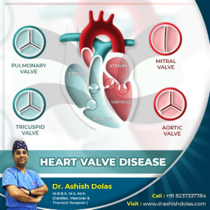
The four heart valves:
- Tricuspid valve: Located between the right atrium and the right ventricle.
- Pulmonary valve: Located between the right ventricle and the pulmonary artery.
- Mitral valve: Located between the left atrium and the left ventricle.
- Aortic valve: Located between the left ventricle and the aorta.
Heart valves can have one of two malfunctions:
Regurgitation
The valve(s) does not close completely, causing the blood to flow backward instead of forward through the valve.
Stenosis
The valve(s) opening becomes narrowed or does not form properly, inhibiting the flow of blood out of the ventricle or atria. The heart is forced to pump blood with increased force in order to move blood through the stiff (stenotic) valve(s).
Heart valves can have both malfunctions at the same time (regurgitation and stenosis). When heart valves fail to open and close properly, the implications for the heart can be serious, possibly hampering the heart’s ability to pump blood adequately through the body. Heart valve problems are one cause of heart failure.
Causes
- Energy changes in the body weaken the valve tissue. This is called myxomatous degeneration. It happens most often in elderly patients and commonly affects the mitral valve.
- A buildup of calcium on the aortic or mitral valves causes the valves to thicken. This is called calcific degeneration.
- An irregularly shaped aortic valve or a narrowed mitral valve is usually a congenital defect, which means people got it by birth.
- Use of the anti-obesity medicines fen-phen and Redux, which were removed from the market after being linked to heart valve disease.
- An infection in the lining of the heart’s walls and valves (the endocardium). This is called infective endocarditis.
- Coronary artery disease.
- Heart attack.
Symptoms
Symptoms depend on the patient and the type and severity of valve disease. Many times, patients have no symptoms at all. In other cases, valve disease may take its damage over many years. In time, patients may develop congestive heart failure. Also, valve disease may lead to heart muscle disease (cardiomyopathy), an irregular heartbeat (arrhythmia) and blood clots.
Diagnosis
Only a doctor can tell if one has valve disease by listening the heart with a stethoscope. Doctors can listen for the distinct clicking sounds or murmurs of valve disease. The doctor may order include other tests, like
- A chest x-ray, which can show if your heart is enlarged.
- Echocardiography, which can show the thickness of your heart’s walls, your valves’ shape and action, and the size of your valve openings. Doppler echocardiography (ultrasound) can be used to find out how severe the narrowing (stenosis) or backflow (regurgitation) of blood is.
- Electrocardiography (EKG or ECG) can be used to find out if your ventricles or atria are enlarged. This test can also determine if you have an irregular heartbeat (arrhythmia).
- Coronary angiography it lets doctors see your heart as it is pumping. Angiography can help identify a narrowed valve or any backflow of blood. For more information